
Monday, September 26, 2011


Saturday, September 17, 2011
3.2 Fertilisation
Key points:
1. The process begins with the adult male and the adult female. The cells which make up an adult have a complete set of chromosomes. Diploid means that they have a complete set of chromosomes. These cells divide in the testis to produce a half set and in males this is the sperm cell, and in females its the egg. This is known as meiosis, and this will half the number of chromosomes. From 2n -> n. So 23 chromosomes.
2. In sexual reproduction these two cells are brought together and they are joined together so that it forms just one cell. This process is known as fertilisation. It involes combining two half sets into one diploid. 23 from sperm + 23 from egg = 46 known as zygote.
3. This new cell is a combination of the male and female chromosomes. This goes through a process known as mitosis, where the cells will divide. One cell -> Two cells. They will both contain 46 chromosomes, and they will divide and the cell divisions will also have 46 chromosomes. All will contain 2n diploid number of chromosomes. Once there are a group of divided cells, this structure will be known as an embryo.
3.9b Female reproductive system
Key Points:
1. Before pregnancy occurs, the uterus structure is no larger than an orange.
2. The ovary is where meiosis occurs and where the eggs are made.
3. The oviducts carry the eggs to the uterus, but also the location where fertilisation may take place.
4. The uterus is in the centre, and the uterus wall is made of muscle and will stretch to accomadate pregnancy. The lining of the uterus accepts and develops the fertilised egg and would develop into an embryo. The development of the placenta would be seen here.
5. Sperm cells enter the uterus through the cervix which is the entrance to the uterus. Uterus space is where the sperm cells move and egg cells move, and its better known for where the baby develops.
6. The vagina is where the penis is introduced and the vagina collects the sperm cells and allows them to pass through the cervix into the uterus.
3.9a Male reproductive system
Key Points:
1. The function of the bladder is to store urine.
2. The function of the testis is to carry out meiosis that produces the gamete called a sperm cell. Sperm cells are stored in the epididymis.
3. The vas deferens carries sperm cells to the penis during sexual stimulation. The walls of this tube pulse.
4. The prostate adds 20-30% of the volume of semen and contains sugars and its also alkaline. The alkaline is thought to neutralise the acidic secretions of the vagina.
5. The seminal vesicles produce sugar based secretions, and they're also alkaline. They make up 70% of the semen.
6. When sperm cells are combined with the prostate and seminal vesicles then it is called semen, and this is carried through a tube into the urethra. The urethra is the common tube that joins the left and right testis. Takes semen down the penis, and is also the exit for urine.
7. The function of the penis is to carry the sperm cells into the vagina during sexual intercourse.
Saturday, September 10, 2011
3.12 Amniotic Fluid
Key Points:
In the diagram, surrounding the rest of the embryo is a fluid. That fluid is called the amniotic fluid. One function of the amniotic fluid is that it protects the embryo, the fluid which is largely water, cannot be compressed. If the fluid was squeezed, it would absorb the pressure. Any blows or hits or force applied to the uterus wall will be nullified because the amniotic fluid absorbs the pressure and prevents the embryo from getting damaged.
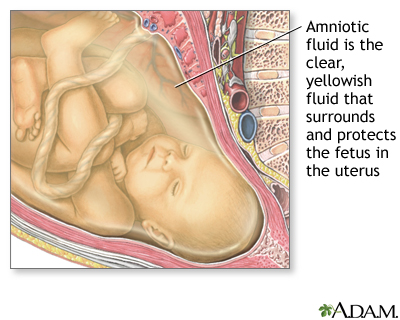 |
| http://www.nlm.nih.gov/medlineplus/ency/imagepages/17008.htm |
3.11 Placenta
Key Points:
1. The structure is known as the uterus, and on one side of the uterus there is a structure known as the placenta. The role of the placenta. When the child is in the uterus, its a water filled environment, amniotic fluids, at this time child cannot breathe or digest. Also unable to properly carry out excretion. So how does the child obtain nutrition?
2. Growing out of the embryo is the placental structure, there's something known as the umbilical cord. In the diagram, the embryo would be at the end of the cord. There are blood vessels on the umbilical cord and these blood vessels lead from the embryo to the placenta and they spread out to form a structure known as the placenta.
3. The placenta biological grows out of the developing embryo, NOT the mother. The blood vessels are the childs blood vessels.
4. The structure on the side is the lining of the uterus. The placenta grows into the wall of the uterus. In the blood stream of the mother there would be stuff like glucose, fats and amino acids. These would travel through her blood stream and into her uterus, and they'll cross into the childs blood at the placenta.
They go from the mothers blood to the childs blood through the placenta. To make this efficient the placenta has a LARGE surface area.
Subscribe to:
Posts (Atom)